Microsoft ends support for Internet Explorer on June 16, 2022.
We recommend using one of the browsers listed below.
- Microsoft Edge(Latest version)
- Mozilla Firefox(Latest version)
- Google Chrome(Latest version)
- Apple Safari(Latest version)
Please contact your browser provider for download and installation instructions.
May 24, 2023
NTT Corporation
Long-term functional measurements from modular 3D neuronal networks using rolled-up 3D electrode array
~Functional characterization of brain-mimicking culture by bioelectronics~
NTT Corporation (NTT; head office: Chiyoda-ku, Tokyo; President and CEO: Akira Shimada) has succeeded in the long-term measurement of neural activity from a neural network (*1) of connected three-dimensional (3D) neuronal aggregates (*2) using NTT's original method (*3) for deforming planar graphene (*4) into 3D graphene.
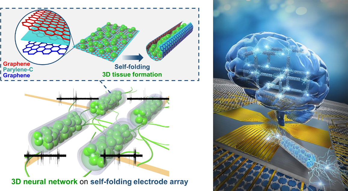
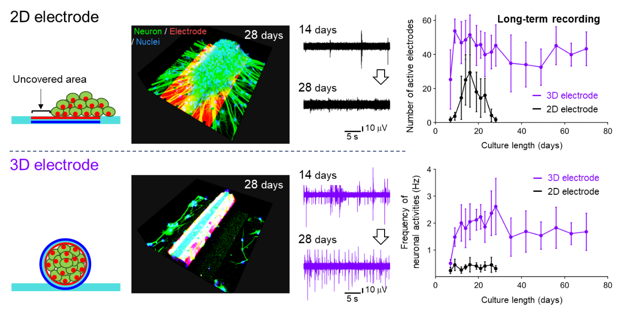
In this study, NTT developed a 3D graphene-based roll that works as a cell scaffold (*5) for creating a 3D neuronal aggregate and then utilized the graphene surface as an electrode for measuring neural activities. This enables us to measure neural activities for 70 days from a neural network of interconnected 3D neuronal aggregates. The recordings show that the obtained spatiotemporal pattern of neural activity (*6) is similar to the activity pattern in the brain's network, which exhibits a mixture of synchronous and asynchronous activity.
This platform will be utilized as a powerful tool to examine neural functions in brain-mimicking cultures (networks of 3D neuronal aggregates) and thereby help us to establish brain-on-a-chip technology to accelerate research related to brain development, disease, and drug discovery.
The details of this achievement were published in the American scientific journal Advanced Functional Materials on May 1, 2023 (U.S. Eastern Time).
 Figure 1. (Left) 3D electrode array for measuring activities from 3D neuronal network,
(Right) an image of brain-on-a-chip.
Background
The brain is an organ that functions as a network of neurons. The mechanisms underlying how neurons assemble, grow, and function remain unclear, so in recent years, brain-on-a-chip technology has been attracting attention as a means of clarifying these mechanisms by assembling cultured neurons and measuring the function of neuronal assembly. Tissue engineering technologies (*7) have been developed to create 3D neuronal aggregate by providing 3D scaffolds for neuronal culture, and there is a recently developed method that measures neural activity from 3D neuronal aggregate using 3D neural interfaces (*8). Although the neuronal network in the human brain consists of multiple neuronal assemblies, it is challenging to apply this measuring method to multiple 3D neuronal aggregates due to the technical difficulty of aligning electrodes on each neuronal aggregate independently.
We have developed an original method to fold graphene into a cylindrical shape by manipulating its flexibility and utilized the cylindrical shape to create a 3D neuronal aggregate. Additionally, many studies have used graphene for neuronal interfaces due to its high electroconductivity. Furthermore, its biocompatibility (*9) and transparency facilitate the optical observation of cell growth. Therefore, foldable graphene is expected to work as both a cell scaffold for 3D neuronal aggregate and as a recording electrode.
Achievements
In this study, we demonstrate that the 3D graphene electrode enables us to measure the neuronal activities from 3D neuronal aggregates grown inside graphene (Fig. 1). Since the 3D neuronal aggregate is formed inside the cylindrical 3D electrode, it is easy to form a pair of the 3D neuronal aggregate and the electrode, resulting in a stable maintenance of cell-electrode contact. Neural activities can be measured over a period of 70 days, and the frequency of activities is three times more than when using a flat electrode (Fig. 2).
 Figure 2. Long-term measurements from 3D neuronal aggregates.
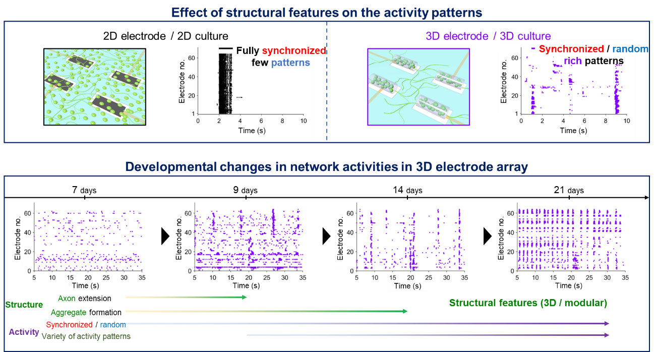
An 8×8 array of 3D graphene electrodes (*10) can measure activities independently from multiple 3D neuronal aggregates corresponding to each electrode. This recording capability allows for activity measurement from a neural network that consists of interconnected 3D aggregates (Fig. 1). When conventional planar electrodes are used, cultured neurons form a planar and uniform network, and exhibit fully synchronized activity (*11) between neurons. In contrast, the network of interconnected 3D aggregates exhibits various spatiotemporal patterns (*12) of neuronal activity with a mixture of synchronous and asynchronous activity. Additionally, these patterns alter along the culture length. In the human brain, the modular structure (*13) of cellular clusters contributes to the generation of diverse activity patterns. In this study, by combining the cell scaffold for forming 3D aggregates and signal measurement, we reveal that the cultured neuronal network with structural characteristics such as three-dimensionality and modularity exhibits activity patterns similar to those in the human brain. Further, we demonstrate that the long-term recording from the neuronal network enables us to evaluate developmental changes in the characteristic activity patterns (Fig. 3). Therefore, we believe that the functional investigation of brain-mimicking neuronal networks will help us better understand the functions of brain neuronal networks in physiological and pathological conditions.
 Figure 3. Evaluation of network dynamics and its developmental changes.
Technical features
<Technical feature 1: Combination of cell scaffold with electrode>
Our studies on conductive biomaterials have enabled us to successfully combine the cell scaffold and the electrode by utilizing the high electroconductivity and biocompatibility of graphene. The deformable graphene-based film consists of graphene and parylene-C (*14), a polymer (Fig. 1). Thanks to the biocompatibility of both materials, the process of film deformation and cell encapsulation don't affect the cell viability. Although it is difficult for the previous method to make graphene the inner surface of a cylindrical shape, we modify the method by sandwiching the parylene-C layer between two layers of graphene to make the graphene form the inner electrode surface.
<Technical feature 2: Multisite and long-term measurement for investigating the functional development of 3D neuronal network>
The maintenance of 3D aggregate inside the folded electrode allows for the stable measurement of neuronal activities from individual 3D aggregates. The multi-site recordings make it possible to obtain the spatial patterns of neuronal activities representing how aggregates are interconnected. Additionally, the capability of long-term measurement enables us to investigate how the interconnection changes with neuronal development progresses.
Perspective
To investigate the detailed functions of a neuronal network, the pattern of electrodes should be varied. For example, placing multiple electrodes on each aggregate will enable the evaluation of the spatial distribution of neuronal activities in each aggregate. Additionally, co-culturing the various cell types such as glial cells and neurons in other brain regions will emulate different brain's structural features. Our method recapitulates the structural and functional characteristics of the neuronal network including modularity and three-dimensionality and can thus be utilized to help us better understand the functions of the brain and to investigate drug safety and efficacy in the brain-mimicking environment.
Paper information
Authors:K. Sakai, T. F. Teshima, T. Goto, H. Nakashima, M. Yamaguchi
Title:Self-Folding Graphene-Based Interface for Brain-Like Modular 3D Tissue
Journal:Advanced Functional Materials
Publication date:May 1, 2023 (U.S. Eastern Time)
Link:https://onlinelibrary.wiley.com/doi/full/10.1002/adfm.202301836
Glossary
*1Neuronal network
Neurons communicate with each other by building synaptic interconnections. Synchronization of the recorded neuronal signals between different electrodes represents the connectivity of neurons.
*2Three-dimensional neuronal aggregate
Neurons grow and form 3D cellular aggregates called spheroids or organoids. The 3D culture is known to mimic the microenvironment in the brain.
*3Three-dimensional self-assembly of graphene-based film
NTT developed an original method to induce the self-folding of graphene by attaching it onto a polymer thin film. The graphene-based film self-folds into a rolled-up structure that can be used as a cellular scaffold.
https://group.ntt/jp/newsrelease/2019/10/18/191018a.html
*4Graphene
Graphene is an atomic layer of carbon and features high electroconductivity, transparency, and biocompatibility. The unique material properties have attracted much attention in the research field of biomedical engineering.
*5Cell scaffold
While neurons are typically cultured on a 2D culture plate, cell culture on 3D scaffolds creates 3D cellular constructs.
*6Neuronal activity
Neurons encode information through an electrical neuronal activity in which the potential of a cell membrane dramatically rises and falls over a period of 1 ms. The recording electrode nearby the neuron senses the series of potential changes as a neuronal activity.
*7Tissue engineering
Tissue engineering is a research field in which researchers create tissues with the required functions and structures by assembling living cells to achieve regenerative medicine and drug discovery.
*8Neural interface
The neural interface records the changes in extracellular potential caused by neuronal activities. Culturing neurons on neuronal electrodes allows for recording from the cultured neurons. Additionally, electrodes inserted inside the body can record signals from animals and humans.
*9Biocompatibility
Biocompatibility refers to the property of having affinity with cells and tissues and not eliciting a reaction to foreign substances in the body.
*10Microelectrode array
Microelectrode array can record neuronal activities from multiple neurons cultured on the substrate. Recording with microelectrode array is less invasive than the patch-clamp recording method in which an electrode is directly inserted into neurons, thus making it suitable for long-term recording.
*11Synchronized activity
An activity in a neuron induces an activity in the other connected neuron. When a neuron group repeatedly exhibits activities at almost the same time, the collective activity is referred to as synchronized activity and is known to propagate from a brain region to different regions. In the case of a cultured neuronal network, many researchers have characterized the synchronized activity to investigate the network functions.
*12Spatiotemporal pattern
Neural activity measured by multiple electrodes contains both spatial information and temporal information: namely, the synchronization of neural activities. The spatiotemporal pattern of activities represents the functional property of the neural network.
*13Modular structure
Units with a certain function are regarded as modules and are combined to work as an organized system. Brain function is achieved by interconnecting different brain regions and cell clusters as modules. In this study, the modular structure is formed by interconnecting the 3D neuronal aggregates that work as individual modules.
*14Parylene-C
Parylene-C is a polymer that consists of chloroparaxylene and has excellent biocompatibility and insulating properties. Since it adheres to graphene via aromatic rings through π-π interactions, the graphene does not detach when the graphene-parylene-C film is folded.
Media Contact
NTT Corporation
Science and Core Technology Laboratory Group, Public Relations
nttrd-pr@ml.ntt.com
Information is current as of the date of issue of the individual press release.
Please be advised that information may be outdated after that point.
NTT STORY
WEB media that thinks about the future with NTT


